Stratham-Newfields Veterinary Hospital
Providing Seacoast Pets a Higher Standard of Care 
  • Home
  • About Us
    • More about AAHA
    • More about ABVP
    • Pet Health Insurance
  • Contact Us
    • Petly information
  • Our Team
    • Doctors >
      • Dr. Dennis
      • Dr. Young
    • Nurses
    • Client Service Representatives
    • Assistants
    • "Alumni"
    • and, of course, you
    • Join Our Team
  • Our Services
    • Dental >
      • Dental X-Rays
    • Surgery
    • Anesthesia Safety
    • Pain Management
    • Diagnostic Imaging
    • Patient Care
    • Integrated Pet Care >
      • Acupuncture
      • PRP for arthritis
    • Emergency and Triage
  • Take a Tour
    • Reception
    • Cat-Friendly Exam Rooms >
      • Cats are not small dogs :)
    • Dog-Friendly Exam Rooms
    • Pharmacy
    • Dentistry >
      • Dental X-Rays
    • Hospitalized Patients >
      • Treatment and ICU
    • Why we are different
    • Surgical Procedures >
      • Human-Grade Surgical Suite
      • Pre-Operative Exam & Labs
      • Patient Monitoring
    • Digital X-Ray and Sonography
    • Boarding Facilities
  • Pet Parent Education
    • Dental care videos and books - free!
    • Painful tooth conditions
    • Tooth abscess
    • Travel & Pets
    • Hip arthritis (cat)
    • Bladder stones
    • Corn cob in stomach (dog)
    • Heart Diseases
    • Ear Care
    • Ear canal mass
    • How to apply eye drops
    • Anal gland issues
    • Thyroid Disease
    • Diabetes mellitus >
      • Diabetes in cats
      • Diabetes in dogs
    • Links to other info
    • The Dog Park

Removal of an ear mass using a video otoscope.
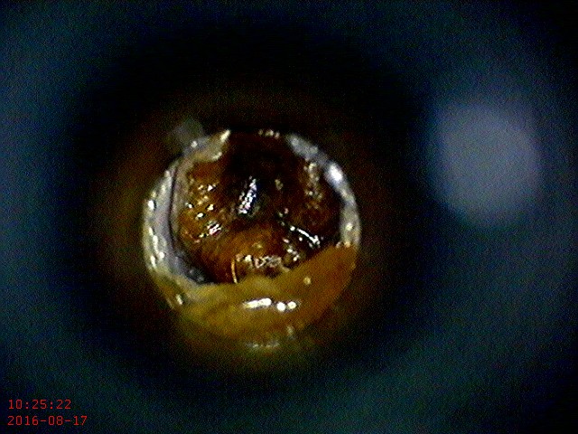

Picture

Before

This is a mass deep in the ear canal of a cat.  It had been non-responsive to medical treatment for a few months, and the cat continued to develop ear infections. 
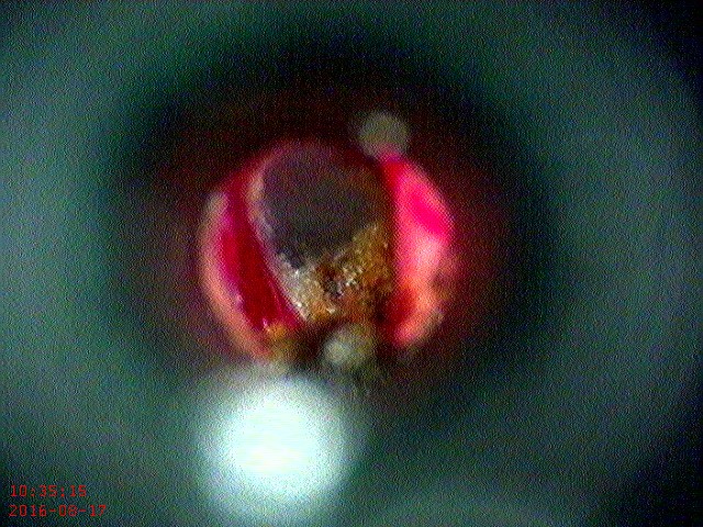
Picture

During

​This is after removal of the mass.  The red is a little blood from where it was attached.  The brown is some remaining wax and debris that was trapped behind it.  The grey area above the brown is the ear drum.
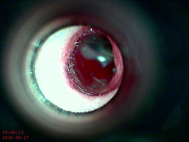
Picture

After

This is after cleaning and removing the remaining wax and infected material.  The red is a blood clot forming.  The translucent white is the ear drum.
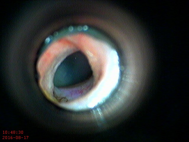
Picture

Normal

​This is the other ear.  Notice the pale pink compared to the red, irritated look of the other side.  The ear drum is translucent. 

 Stratham-Newfields Veterinary Hospital, 8 Main Street, Newfields NH 03856
Phone: 603-778-7687   | Fax: 603-778-2471  |   vip@snvh.net
​Directions and Additional Contact Information
e-mail us
Site powered by Weebly. Managed by IDEXX Laboratories